Proof of weight based enhanced CT scan contrast agent injection protocol
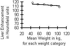
The post-contrast liver enhancement values (in Hounsfield units) are all very similar, across all weight categories.
| For Abdomen scans. Scans that started at the top of the liver. | |||||
| Weight category | 38-47 kg | 48-63 kg | 64-77 kg | 78-97kg | 98-117kg |
| Dose | 70ml | 80ml | 100ml | 120ml | 140ml |
| Liver Hounsfield units | 118.3 | 114.6 | 113.7 | 111.5 | 106.3 |

When this data is graphed, it shows that the lighter patients had slightly brighter liver enhancement than the heavier patients, even though the lighter patients received smaller doses of intravenous contrast agent. If you were expecting the smaller contrast doses to result in lower liver enhancement, I hope you are suitably suprised by this data.
| For Chest scans. Scans that started at the top of the chest. | |||||
| Weight category | 38-47 kg | 48-63 kg | 64-77 kg | 78-97kg | 98-117kg |
| Dose | 70ml | 80ml | 100ml | 120ml | 140ml |
| Liver Hounsfield units | 117.7 | 114.2 | 114.7 | 111.4 | 108.2 |

This data, from scans that began above the chest, shows similar results to the scans that began above the liver. Shown graphed, the lighter patients again had slightly brighter liver enhancement than the heavier patients, even though the lighter patients received lower doses of intravenous contrast agent.
Conclusion: This method works. The practice of dividing patients into weight categories based on their body weight, and giving them a weight-based dose of intravenous contrast agent during CT scanning, results in liver enhancement values that are very similar across all weight categories.
Additional analyses of the supporting study data available at: Hepatic veins and liver enhancement in each weight category, at different scan delay times.
back to weight based CT scan protocol index
End of page Navigation links: Halls.md home or Back to top
Copyright © 1999 - present. Steven B. Halls, MD . 1-780-608-9141 . [email protected]