Construction and Goals of the CT scanning Chest protocol
Use the "Chest" protocol, when the requested scan is only for the chest, (not specifically requesting abdomen or liver).
Scans of the chest always include slices of the superior liver. In an oncology setting, it is not uncommon for unsuspected liver metastases to be discovered on the lowest slices of Chest CT scans. Finding unsuspected liver metastases occurs more frequently than missing significant mediastinal lesions. This is especially true in breast cancer. Furthermore, as part of the natural history of patients with cancer, a patient who initially only needed a CT of the chest, will likely need CT of the abdomen in subsequent months, and when they do, it is helpful to have a comparable previous CT scan. Therefore, we believe (at the Cross Cancer Institute) that the timing of contrast injection for Chest CT scans, should be optimized for liver diagnosis, in preference to optimizing for mediastinal diagnosis (which is relatively unaffected by altering the timing of contrast injection).
The scanning direction is superior-to-inferior, starting one or two slices above the top of the lungs. The speed of the scanner and the slice thickness used, will influence the amount of time it takes to scan down to the top of the liver. When the scanning reaches the liver, it is desirable to have the time elapsed (since injection began) be in the range of 60-70 seconds, which is typically suggested for portal-venous-phase imaging. See the analysis of scan Delay timing, for additional info.
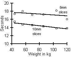
The length of the chest varies slightly with body weight, so the time it takes to scan the chest varies slightly with body weight, but as the tables and graph below show, the effect is not significant. However, slice thickness makes a difference. On our scanner, when 8mm thick slices were used, the average time to scan the chest was 17.9 seconds, compared to 14.3 seconds when 10mm thick slices were used. (Using a spiral scanner with 1.0secs per revolution, 1.5cm per second (1.5 pitch).)
| Time it takes to scan the chest, to the top of the liver. | |||
|---|---|---|---|
| Weight | 8mm | 10mm |  |
| 118 + kg | 16.0sec | 13.8sec | |
| 98 - 117 kg | 18.2sec | 13.8sec | |
| 78 - 97 kg | 17.2sec | 14.3sec | |
| 64 - 77 kg | 17.1sec | 14.6sec | |
| 48 - 63 kg | 18.0sec | 14.7sec | |
| 38 - 47 kg | 17.3sec | 15.4sec | |
| < 38 kg | 13.5sec | ||
When using 8mm thick slices, it takes on average 17.9 seconds to scan the chest, plus or minus a standard deviation of 2.59 seconds. This population standard deviation indicates inter-patient variability, whereby some patients have shorter length chests, and some have longer chests. Knowing that 95% of the population is included within ± 1.96*standard deviation, ie 5.08 seconds variability, the Chest protocol was constructed with 5 seconds "padding".
To put this theory into practice, the Chest protocol scan delays were constructed as follows:
Step 1: Using 8mm slices, it takes about 17.9 seconds to scan the average chest. To accomodate short chests, subtract 5.08 seconds. Thus 12.82 seconds is the minimum time needed to scan the chest.
Step 2. Depends on the weight group. For 64-77 kg, if the goal is to start scanning the top of the liver at a minimum earliest of 62 seconds, subtract 12.82, and start scanning at the top of the chest after 49 seconds delay for injection. What this accomplishes is that even the short-chest patients should start liver scanning at about 62 seconds, and the average patients should start scanning the liver at about 67 seconds.
Step 3. repeat step 2 for each weight category, substituting the desired minimum scan delays to start liver scanning into the formula.
back to the weight based CT scan protocol technique index
End of page Navigation links: Halls.md home or Back to top or
Copyright © 1999 - present. Steven B. Halls, MD . 1-780-608-9141 . [email protected]